
Same Blood Test, Different Answer: MCED and Tumour Markers
Why the value of a cancer blood test is decided less by the assay than by who is sitting in front of it, and what the first randomised trial actually found.
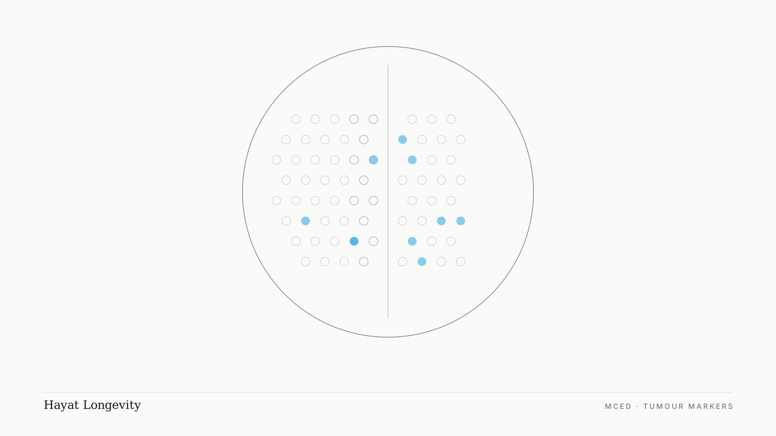
Conventional tumour markers are serum proteins validated for monitoring diagnosed malignancy, not for detecting it in asymptomatic individuals. Multi-cancer early detection (MCED) tests analyse circulating cell-free DNA for methylation patterns shared across many tumour types, and report a binary cancer signal plus a predicted tissue of origin.
MCED specificity is high, reported between 97.4 and 99.6 per cent across published studies, while sensitivity is moderate and lowest in stage I disease. The positive predictive value of any screening test is determined by the underlying prevalence of disease in the population tested, which is why MCED performs meaningfully better in individuals with elevated baseline risk than in the general population.
Neither test class replaces mammography, cervical screening, colorectal screening or low-dose CT.
Key takeaways
- Tumour markers such as CEA, CA 125 and CA 19-9 are validated for monitoring known cancer, not for finding it in people without symptoms.
- MCED tests read methylation patterns in circulating cell-free DNA, reporting a cancer signal and a predicted tissue of origin from one sample.
- Predictive value is a property of the population, not the assay. The same test yields mostly false positives at low prevalence and mostly true positives at high prevalence.
- NHS-Galleri, the first randomised trial, missed its primary endpoint in 2026 but reduced stage IV diagnoses and emergency presentations.
- Sensitivity in stage I disease remains the principal limitation. A negative result does not exclude early cancer.
- No MCED test currently holds FDA approval, and none replaces established screening programmes.
What determines whether a screening test is useful?
Four quantities describe any screening test.
Sensitivity is the proportion of people with disease who test positive. Specificity is the proportion of people without disease who test negative. These are properties of the assay and remain stable across populations.
Positive predictive value (PPV) is the proportion of positive results that represent true disease. Negative predictive value (NPV) is the equivalent for negative results. Neither is a fixed property of the assay. Both vary with disease prevalence in the population being tested.
This relationship is defined by Bayes' theorem:
The practical consequence is central to everything that follows. An identical test, unchanged in every respect, produces mostly false positives in a low-prevalence population and mostly true positives in a high-prevalence one. Test selection is therefore inseparable from patient selection.
Conventional tumour markers
How they work
Tumour markers are macromolecules, predominantly glycoproteins, produced either by tumour cells or by host tissue in response to a tumour. They are quantified in serum by immunoassay and reported against a reference interval.
| Marker | Molecular class | Principal association | Documented non-malignant elevation |
|---|---|---|---|
| CEA | Glycoprotein, oncofetal antigen | Colorectal, some lung and breast | Tobacco smoking, inflammatory bowel disease, cirrhosis, pancreatitis, peptic ulceration |
| CA 19-9 | Sialylated Lewis antigen | Pancreatic, biliary tract | Pancreatitis, cholangitis, any biliary obstruction, cirrhosis, diabetes |
| CA 125 | MUC16 mucin | Epithelial ovarian | Endometriosis, uterine fibroids, menstruation, pregnancy, pelvic inflammatory disease, cirrhosis, congestive cardiac failure, any peritoneal irritation |
| CA 15-3 | MUC1 mucin | Breast | Benign breast disease, hepatic disease, other adenocarcinomas |
| AFP | Oncofetal glycoprotein | Hepatocellular, non-seminomatous germ cell | Viral hepatitis, cirrhosis, pregnancy |
| PSA | Kallikrein-3 serine protease | Prostate | Benign prostatic hyperplasia, prostatitis, urinary retention, recent ejaculation, perineal pressure |
| EBV DNA | Viral cell-free DNA | Nasopharyngeal carcinoma | Recent or reactivated Epstein-Barr virus infection |
What they are validated for
Conventional markers have established evidence for three applications, all of which occur after diagnosis:
- Treatment response assessment. Serial measurement tracks tumour burden during therapy.
- Recurrence surveillance. A rising trend in a previously normalised marker can precede radiological evidence of relapse.
- Diagnostic adjunct. Interpreted alongside imaging and histopathology, not independently.
In each case the marker functions as a longitudinal trend within an individual whose disease status is known.
Why they fail as screening tests
Three distinct failure modes apply.
Analytical specificity is insufficient. As the table shows, every marker in routine use is elevated by common benign conditions. CA 125 is elevated in a substantial proportion of premenopausal women through physiological and benign gynaecological causes alone. CEA is elevated in smokers as a baseline finding.
Sensitivity is stage-dependent and low in early disease. Marker elevation generally correlates with tumour bulk. Stage I malignancies frequently produce values within the reference interval. A separate constraint applies to CA 19-9: individuals with a Lewis-negative phenotype, comprising approximately 5 to 10 per cent of most populations, lack the fucosyltransferase required to synthesise the antigen and cannot produce a detectable result irrespective of disease burden.
Predictive value collapses at low prevalence. Applying the formula above to a marker with 95 per cent specificity and 70 per cent sensitivity, tested in a population with an annual cancer incidence of 0.5 per cent, yields a PPV of approximately 6.6 per cent. Roughly 93 of every 100 positive results would be false. This result is arithmetic, not a defect of any individual assay, and it is the reason major guideline bodies do not recommend unselected tumour marker panels for screening asymptomatic adults.
The exceptions, and why they are exceptions
Where markers are used before diagnosis, it is always within a defined high-risk cohort, precisely because prevalence enrichment restores predictive value.
- AFP with abdominal ultrasonography at six-monthly intervals for hepatocellular carcinoma surveillance in cirrhosis and chronic hepatitis B infection. Hepatitis B carriage is prevalent across Southeast Asia, making this cohort large in this region.
- PSA for prostate cancer, offered through shared decision-making rather than routine screening, reflecting a finely balanced benefit-to-harm ratio.
- Plasma EBV DNA for nasopharyngeal carcinoma in endemic populations, notably southern Chinese ancestral groups across Southeast Asia, where incidence is elevated by an order of magnitude relative to global rates.
Multi-cancer early detection tests
How they work
Cells release fragments of DNA into the circulation during apoptosis and necrosis. In an individual with malignancy, a small fraction of this circulating cell-free DNA (cfDNA) originates from tumour cells and is termed circulating tumour DNA (ctDNA).
Tumour-derived cfDNA carries epigenetic modifications distinguishable from those of normal tissue, principally aberrant patterns of cytosine methylation at CpG dinucleotides. These patterns are both tumour-associated and tissue-specific, since methylation signatures differ systematically between organs.
MCED assays apply targeted bisulphite or enzymatic conversion sequencing across large panels of methylation regions, and classify the resulting patterns using machine learning models trained on tens of thousands of clinical samples. Some platforms incorporate additional analyte classes, including protein biomarkers and fragmentomic features such as cfDNA fragment length distribution.
Output comprises two elements:
1. Cancer signal detected or not detected. A binary classification. 2. Cancer signal origin. Where a signal is present, a ranked prediction of the tissue of origin, which directs the diagnostic pathway rather than prompting undirected whole-body imaging.
The American Cancer Society describes these tests as examining blood or other body fluid for DNA, RNA or protein fragments from abnormal cells, and states that a positive result requires further testing to establish whether cancer is present, its type and its location.
Why the category exists
Population screening programmes address a small number of malignancies: breast, cervix, colorectum, and lung in eligible smokers. GRAIL cites data indicating that approximately 7 in 10 cancer deaths arise from tumour types with no recommended screening pathway, including pancreatic, hepatic, ovarian and oesophageal carcinoma. These are predominantly diagnosed symptomatically at advanced stage.
What the trials found
NHS-Galleri. The first and only randomised controlled trial of an MCED test at population scale. 142,250 participants aged 50 to 77 were enrolled in England, randomised 1:1 to annual MCED testing or standard care, tested at up to three annual visits. Primary results were presented at the ASCO Annual Meeting in May 2026.
The primary endpoint, a reduction in stage III or IV cancer incidence, was not met. Across three screening rounds the incidence rate ratio was 1.03 (95 per cent CI 0.92 to 1.14, p = 0.6324) in the pre-specified group of twelve cancer types. In absolute terms, 706 stage III or IV cancers were diagnosed in the intervention arm against 688 in the control arm.
The secondary findings are where the trial becomes interesting. Stage IV diagnoses of those twelve cancers fell by approximately 14 per cent overall, and the effect strengthened with successive rounds: 9 per cent in the first, 22 per cent in the second, 26 per cent in the third. Stage I and II diagnoses rose by 16 per cent. Screen-detected cancers increased roughly four-fold, and cancers presenting clinically through symptoms or as emergencies fell by around 25 per cent, which matters because those routes carry the worst outcomes.
Expert commentators quoted by the UK Science Media Centre were divided rather than unanimous. The substantive criticisms were that the trial provides no basis yet for population-scale implementation, and that implementation would divert finite CT and biopsy capacity away from symptomatic patients. Others noted that the trial was, by necessity, a compromise: a mortality endpoint would have required close to two decades, by which time the technology would likely be obsolete. Mortality data are expected in approximately two years, and follow-up has been extended.
PATHFINDER 2. The full cohort of 35,878 adults aged 50 and over reported at ASCO in 2026. Adding the MCED test to standard screening increased cancer detection up to 6.5-fold, at a specificity of 99.6 per cent, equating to a false positive rate below 0.4 per cent. Positive predictive value was 60.3 per cent, and episode sensitivity was 69.8 per cent for the twelve cancers responsible for around two-thirds of US cancer deaths. More than half of the cancers the test detected were early stage.
Cancerguard. The manufacturer reports 68 per cent sensitivity for the six malignancies with lowest five-year survival, comprising pancreatic, lung, hepatic, oesophageal, gastric and ovarian, at a specificity of 97.4 per cent.
Regulatory status
No MCED test currently holds FDA approval. Both leading tests are available in the United States through laboratory-developed test pathways rather than as approved screening programmes. GRAIL has been pursuing premarket approval for Galleri under a Breakthrough Device Designation, and a decision has been anticipated during 2026, so this position may change.
MD Anderson, Fred Hutch, Dana-Farber and the American Cancer Society state consistently that these assays are not diagnostic, that positive results require confirmatory investigation, and that they do not substitute for recommended screening modalities.
The two classes side by side
| Conventional tumour markers | MCED | |
|---|---|---|
| Analyte | Serum glycoproteins and antigens | Circulating cell-free DNA methylation patterns, with protein or fragmentomic features on some platforms |
| Validated indication | Monitoring diagnosed malignancy | Under investigation for screening asymptomatic individuals |
| Coverage per test | One marker corresponds to one or two tumour types | Multiple tumour types from a single sample |
| Reported specificity | Variable and frequently low in asymptomatic populations | 97.4 to 99.6 per cent in published studies |
| Sensitivity profile | Correlates with tumour burden, low in stage I | Moderate overall, lowest in stage I |
| Output | Continuous value against a reference interval | Binary signal plus predicted tissue of origin |
| Randomised mortality evidence | Not established for screening use | Not yet available |
| Substitutes for standard screening | No | No |
Why who is tested decides what a result means
The opening section established that predictive value is a function of prevalence. This is the scientific basis for restricting MCED to individuals with elevated baseline risk.
Applying the Bayesian formula to illustrative parameters of 99.5 per cent specificity and 50 per cent sensitivity:
| Annual cancer incidence in the tested cohort | Approximate PPV |
|---|---|
| 0.5 per cent (general population, sixth decade) | 33 per cent |
| 2 per cent (enriched risk) | 67 per cent |
| 5 per cent (substantially enriched risk) | 84 per cent |
The assay is identical in all three rows. Only the population changes. Directing testing toward higher-prevalence cohorts increases the proportion of positive results that represent genuine disease, and correspondingly reduces the number of individuals subjected to unnecessary diagnostic investigation.
What raises baseline risk
- Documented pathogenic germline variants, including BRCA1, BRCA2, and the mismatch repair genes associated with Lynch syndrome
- Significant family history, particularly first-degree relatives with malignancy diagnosed at young age, or multiple affected relatives within one lineage
- Chronic hepatitis B or C infection, or established cirrhosis, conferring elevated hepatocellular carcinoma risk
- Substantial cumulative tobacco exposure, current or former
- Advancing age, the single largest non-modifiable determinant of cancer incidence
- Chronic inflammatory conditions with recognised malignant potential, including inflammatory bowel disease and Barrett's oesophagus
- Prior malignancy, noting that several MCED providers exclude individuals with a personal cancer history from their stated indications
What risk enrichment does not fix
Prevalence enrichment improves PPV. It does not alter sensitivity. Two limitations therefore remain unchanged in high-risk cohorts:
Stage I sensitivity remains the principal constraint. Small tumours shed proportionately less ctDNA, and detection probability falls accordingly. A negative result does not exclude early-stage malignancy.
Randomised trial evidence derives from average-risk populations. NHS-Galleri and PATHFINDER 2 enrolled unselected participants by age, not by risk enrichment. The Bayesian argument for high-risk targeting is mathematically sound, but has not itself been tested in a randomised trial with mortality endpoints. Established surveillance protocols for defined high-risk groups, such as six-monthly ultrasonography with AFP in cirrhosis, retain their evidence base and are not displaced by MCED.
Conclusion
Conventional tumour markers are validated for monitoring diagnosed malignancy and demonstrate inadequate predictive value when applied to unselected asymptomatic populations, a limitation arising from the interaction of imperfect specificity with low disease prevalence.
MCED represents a distinct analytical approach with substantially higher specificity, addressing tumour types for which no screening pathway currently exists. Its principal limitation is sensitivity in stage I disease, and randomised mortality evidence is not yet available.
Because predictive value scales with prevalence, the scientific case for MCED is strongest in individuals with defined risk-elevating characteristics. Determining whether an individual falls into such a cohort requires structured risk assessment, including personal history, family history, infection status and exposure history, undertaken with a doctor before any test is selected.
That assessment is the part worth doing first, and it costs nothing but a conversation.
To restate the declaration above: we offer this test, which gives us a commercial interest in your reading this article favourably. The honest position is that MCED is not appropriate for most people who ask about it. If your risk profile is unremarkable, the arithmetic in this article argues against testing you, and that is what we will tell you. Where it is appropriate, it belongs alongside your existing screening, never in place of it.
Our pre-consultation questionnaire covers personal and family history, which is what determines whether any of this applies to you. If you want the wider context on what routine screening does and does not cover, our article on what standard medical checkups miss is a useful companion piece.
Written by Dr. Daniel Chong, Human Performance and Longevity. AOKLINIK Penang.
General information only, not individual medical advice, and not a recommendation to undergo any test. Trial data and regulatory positions in this field are changing quickly; figures are accurate as at August 2026. Whether any screening test is appropriate for you depends on your personal and family history and should be decided with a doctor who knows both.
Frequently asked questions
Can a blood test detect all cancer types?
No. Not all malignancies shed detectable cell-free DNA into the circulation, and no assay covers all tumour types. False positive and false negative results occur with all MCED platforms.
How does MCED differ analytically from a tumour marker panel?
Tumour markers quantify specific serum proteins associated with particular organs. MCED analyses methylation patterns in circulating cell-free DNA that are common across many tumour types and simultaneously tissue-specific, enabling both detection and origin prediction from a single sample.
Does a negative MCED result exclude cancer?
No. Sensitivity is moderate and lowest in stage I disease. A negative result does not modify the indication for mammography, cervical screening, colorectal screening or low-dose CT.
What follows a positive MCED result?
A positive result is not a diagnosis. It initiates a diagnostic pathway, typically imaging directed by the predicted tissue of origin, followed by tissue biopsy where indicated. In PATHFINDER 2, around 60 per cent of participants with a positive result received a cancer diagnosis.
Did the NHS-Galleri trial succeed or fail?
Both readings are oversimplified. The trial missed its primary endpoint: there was no significant reduction in combined stage III and IV cancers, with an incidence rate ratio of 1.03. It did reduce stage IV diagnoses by around 14 per cent, with the effect growing across successive screening rounds, increased stage I and II diagnoses by 16 per cent, and cut cancers presenting as emergencies by around 25 per cent. Mortality data are not yet available.
Why is MCED considered more appropriate for high-risk individuals?
Positive predictive value rises with disease prevalence in the tested population. Testing cohorts with elevated baseline risk increases the proportion of positive results representing true disease and reduces unnecessary investigation, without any change to the assay itself.
Are MCED tests regulator-approved?
No MCED test currently holds FDA approval. Availability is through laboratory-developed test pathways and private providers rather than approved population screening programmes, though regulatory submissions are in progress and this position may change.
Does Hayat Longevity offer MCED testing?
Yes, delivered at AOKLINIK Penang, our licensed medical clinic. That is a commercial interest and we should state it plainly. It is also why this article leads with the primary endpoint that the first randomised trial did not meet. MCED is not appropriate for most people who enquire about it: the case rests on elevated baseline risk, and where risk is unremarkable the arithmetic argues against testing. Any discussion should begin with structured risk assessment rather than with the test.
References
- NHS-Galleri: primary results from a randomised controlled trial to assess the clinical utility of a multi-cancer early detection test in population screening. Journal of Clinical Oncology, 2026 (ASCO Annual Meeting, Abstract LBA100).
- Rosenfeld N. Outcomes from the NHS-Galleri trial: the good, the bad, and the uncertain. BJC Reports, 2026.
- PATHFINDER 2 full cohort performance and safety results. Presented at the ASCO Annual Meeting, 2026.
- Schrag D, et al. Blood-based tests for multicancer early detection (PATHFINDER): a prospective cohort study. The Lancet, 2023.
- Klein EA, et al. Clinical validation of a targeted methylation-based multi-cancer early detection test. Annals of Oncology, 2021.
- Science Media Centre. Expert reaction to full results of the NHS-Galleri trial presented at ASCO, May 2026.
- American Cancer Society. Multi-cancer early detection tests: what to know.